


Understanding Infinity Corrected Objective Resolving Power and Magnification
Key Terminology | Low Magnification Imaging | High Magnification Imaging
In the optics industry, microscopes are used for both machine vision and life science, or biological, applications. Machine vision applications support semiconductor, electronics, assembly, and manufacturing markets, to name a few. Life science applications look at cellular or biological samples at various objective magnifications. To understand the interplay between resolving power, magnification, and other common objective specifications for life science, consider how an infinity corrected objective images skin culture at 5X, 10X, 20X, and 50X magnifications.
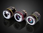
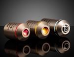
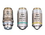
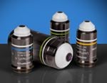
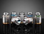
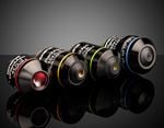
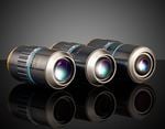
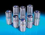
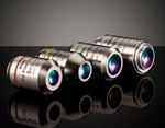
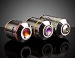
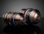
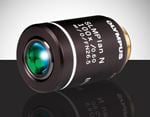
The four application examples detailed in the following sections utilize Edmund Optics infinity corrected objectives. Mitutoyo objectives can be substituted as well. Although Mitutoyo objectives are synonymous with machine vision and industrial inspection, they perform extremely well in low light conditions and applications that focus on cellular and micro inspection.
KEY OBJECTIVE TERMINOLOGY
Infinity corrected objectives image well, no matter the magnification. They are ideal for applications requiring high precision and the modularity of adding optical filters, polarizers, beamsplitters, and in-line illumination components into the optical path. However, there are tradeoffs for using infinity-corrected objectives:
- Higher magnifications yield higher numerical apertures, but shorter working distances and smaller fields of view.
- Lower magnifications yield lower numerical apertures, but longer working distances and larger fields of view.
- Resolution is dictated by magnification, and both increase proportionally to each other.
Magnification, numerical aperture, working distance, and resolution are all related for infinity corrected objectives. Magnification is calculated by dividing the focal length of the tube lens by the focal length of the objective. Numerical aperture (NA) is a function of the focal length of the entrance pupil diameter; NA affects the amount of light entering the infinity corrected system. Working distance (WD) is determined by the parfocal distance of the objective’s optical path; WD is specified as the distance from the front optical element to the object under inspection. Resolving power is one of the trickiest specifications to properly explain. Since it is difficult to visualize what an actual object under inspection will look like at a particular magnification and how to quantify resolving power, it is best to learn by studying the application examples in the following sections. For additional information on key objective terminology, view Understanding Microscopes and Objectives.
APPLICATION EXAMPLES
There are a large number of applications for infinity corrected objective systems. In terms of biological applications, the most common is fluorescence microscopy, ranging from the most basic fluorophore detection systems to the more elaborate confocal and multiphoton fluorescence systems. The most complex of these systems involve high magnifications, precision mechanics, high-quality optical filters, and powerful illumination sources, such as lasers. In contrast, the simpler systems involve a standard broadband light source, basic filtering, simple mechanics, and low to high magnifications depending on the samples under inspection.
Low Magnification Imaging of Skin Culture
Depending on the sample, there are a few rules of thumb to help with the selection of an infinity corrected objective. A typical cell has a size of 10μm; a low magnification and low resolution objective is suitable for imaging a grouping of cells. If one needs to differentiate cellular membranes or intracellular components such as mitochondria, ribosomes, or a nucleus, then resolution on the order of 1μm or less is best.
In Figures 1 – 4, the samples for inspection are 3D Skin Culture Models with a Trichrome Stain, which was cultured and prepared by Zen-Bio Incorporated located in Research Triangle Park, North Carolina, US. In Figure 1, it is clear to see cellular components surrounded by the extracellular matrix (ECM), which holds the tissue together. Within the ECM are the interstitial matrix and basement membrane, where polysaccharides and fibrous proteins reside and act as a compression buffer against external stress. Within the basement membrane reside a number of sheets that are stacked upon one another with epithelial cells resting between. To clearly see the polysaccharide gel and epithelial cells, it is best to use a high magnification infinity corrected objective. The tissue matrix is the surrounding blue stained material, the cells and cellular membrane are marked by the purple stains, and within each cell is a smaller white and partially red stained region that marks denser intracellular material, such as mitochondria and the nucleus.
Figure 1 was captured using #59-876 5X M Plan Apo Objective. This infinity corrected objective has a numerical aperture of 0.14, field of view on a ½" sensor of 1.28mm x 0.96mm, and resolving power of 2μm. Since typical human cells are roughly 10μm in size, the objective specifications for #59-876 make it the ideal choice.
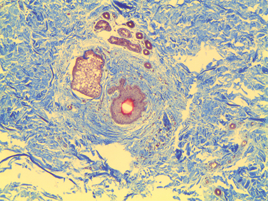
Figure 1: Trichrome Stain of Dermal Tissue Samples at 5X Magnification Using #59-876 Objective
Figure 2 was captured using #59-877 10X M Plan Apo Objective. It has a numerical aperture of 0.28, field of view on a ½" sensor of 0.64mm x 0.48mm, and a resolving power of 1μm. In this image, it is clear to see the stacking and weaving of the ECM and interstitial structures. Additionally, the cellular membrane is very pronounced and it is evident that there are a number of intracellular structures such as ribosomes, mitochondria, and a large nucleus present at the central position.
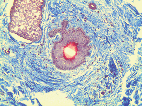
Figure 2: Trichrome Stain of Dermal Tissue Samples at 10X Magnification Using #59-877 Objective
High Magnification Imaging of Skin Culture
Figure 3 was captured using #59-878 20X M Plan Apo Objective. This infinity corrected objective has a numerical aperture of 0.42, field of view on a ½" sensor of 0.32mm x 0.24mm, and a resolving power of 0.7μm. Figure 3 displays the entire cell within the given field of view; the surrounding extracellular matrix is sectioned in greater detail, and the intracellular molecules are much larger and more visible than in either Figure 1 or Figure 2.
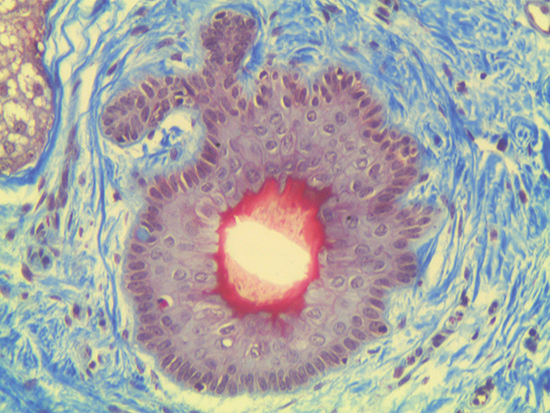
Figure 3: Trichrome Stain of Dermal Tissue Samples at 20X Magnification Using #59-878 Objective
Figure 4 demonstrates 50X magnification - the highest magnification easily achieved without mechanical stages or piezo actuators stabilizing the infinity corrected objective and image plane. At this magnification, slight vibrations from an illuminator or computer’s fan can cause the video feed to dramatically shake and jump out of focus. Figure 4 was captured using #59-879 50X M Plan Apo Objective. It has a numerical aperture of 0.55, field of view on a ½" sensor of 0.128mm x 0.096mm, and a resolving power of 0.5μm. The depth of focus for this particular objective is only 0.9μm, making focusing a tedious process if the appropriate mechanics are not utilized. In Figure 4, the cellular membrane and intracellular components are very clear and vibrant compared to the previous 5X, 10X, and 20X images. Also, the size and shape of the cellular constituents become truly evident. When comparing Figure 1 (5X magnification) to Figure 4 (50X magnification), the increase in magnification is immediately apparent. The resolving power increases by four and the field of view is minimized by a factor of twenty. When imaging at 50X magnification, high light intensity and contrast is required to increase illumination and to digitally adjust shutter speed and gain. The digital settings can be set to automatically compensate for darkness or frame rate – excellent for constructing a digital video microscope for the first time. For additional details on constructing one with off-the-shelf components, please read Digital Video Microscope Objective Setups.
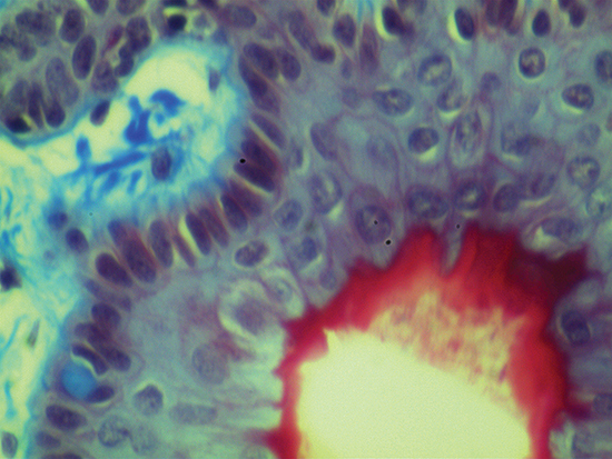
Figure 4: Trichrome Stain of Dermal Tissue Samples at 50X Magnification Using #59-879 Objective
Infinity corrected objectives are ideal for machine vision inspection and life science applications. When imaging biological samples such as dermal tissue, it is important to understand what can be achieved with different objective magnifications. 5X and 10X objectives are ideal for seeing groups of cells and slight structures in the extracellular matrix. 20X and 50X objectives provide greater resolution and are capable of seeing intracellular molecules.
or view regional numbers
QUOTE TOOL
enter stock numbers to begin
Copyright 2023, Edmund Optics Inc., 14F., No.83, Sec. 4, Wenxin Road, Beitun District , Taichung City 406, Taiwan (R.O.C.)
California Consumer Privacy Act (CCPA): Do Not Sell My Information