


Optical Microscopy Application: Phase Contrast
作者: Stephan Briggs
Phase contrast was first utilized and described in 1934 by Frits Zernike. This optical microscopy technique enhances the contrast of transparent specimens, yielding high-contrast images of living cells, microorganisms, and other samples. The main advantage of the phase contrast technique is that living cells and tissues do not need to be killed, fixed, stained, or prepared in any way and can, in turn, be examined in their natural state. Analyzing and recording the dynamics of intricate biological processes becomes very easy with phase contrast optical microscopy.
Image Appearance
A typical phase contrast image has a neutral background and surrounding with varying contrast where light is altered by the specimen (Figure 1). Two very common effects seen in a phase contrast image are halo and shade-off patterns. These occur when the infinite-conjugate focal point does not match for the specimen and background. Although these are common and expected in phase contrast images, they diminish the appearances of details. In general, a bright phase contrast halo is typically visible at a boundary between strong and weak specimen features. These halos are evident due to the circular phase-retarding rings. Specialized objectives, known as apodizing phase contrast objectives, are manufactured to reduce this phenomenon.
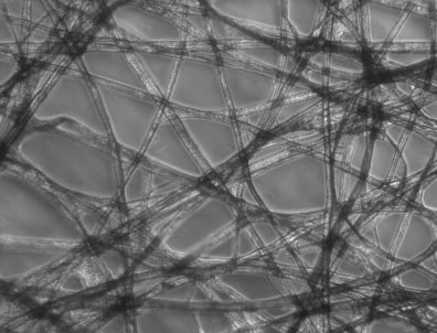
Figure 1: Phase Contrast Image of Tissue Paper
Technical Details
The phase contrast technique translates extremely tiny variations in phase into a noticeable and corresponding amplitude change, and is evident in the difference of contrast in Figure 1. The most important concept of the phase contrast microscope design is the isolation of wavefronts, both surround (undiffracted) and diffracted, that arise from the specimen. To differentiate intensity profiles between a specimen and its surroundings, the undeviated light must be reduced and the phase retarded by a quarter-wave retardance. A brightfield illumination microscope can be upgraded to a brightfield-phase microscope with the introduction of two components to the optical train. For additional information on the brightfield technique, please read Optical Microscopy Application: Brightfield Illumination.
- Phase Plate: mounted on the rear of the objective, this plate selectively alters the phase and amplitude of the light passing through the specimen. Often, microscope objectives have a small ring etched into one of their glass elements to eliminate the need for additional elements.
- Condenser Annulus: opaque flat-black plate with an annular ring that is transparent, and positioned in the front focal plane. This results in the specimen being illuminated by a defocused, parallel light ray.
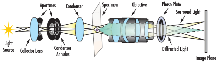
Figure 2: Optical Path of Phase Contrast Microscope
Additional optical microscopy applications include brightfield illumination, darkfield illumination, fluorescence, and differential interference contrast.


or view regional numbers
QUOTE TOOL
enter stock numbers to begin
Copyright 2023, Edmund Optics Inc., 14F., No.83, Sec. 4, Wenxin Road, Beitun District , Taichung City 406, Taiwan (R.O.C.)
Privacy Policy | Cookie Policy | Terms & Conditions | Accessibility
California Consumer Privacy Act (CCPA): Do Not Sell My Information
The FUTURE Depends On Optics®